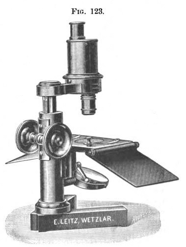
The first application of this erecting system to a Leitz dissecting microscope was reported in the Journal of the Royal Microscopical Society, 1900. It was incorporated into the microscope designed by Richard Friedrich Johannes Pfeiffer (1858-1945). An extract from the Journal is reproduced below:
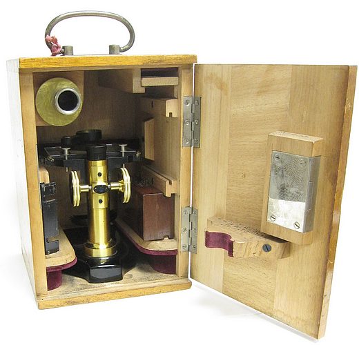
Pfeiffer's New Preparation Microscope.This has been designed by Prof. Pfeiffer to meet a want felt by him in his work on malarial parasites. He considered that an instrument of this kind should give an erect image, a maximum object-distance, large field, and sufficient magnification. Fig. 123 shows the Microscope, which is built on the ordinary principle, but the inverted image produced by the objective is by a suitable prism arrangement presented to the eye as erect. These prisms are contained in a somewhat wide brass tube, which on its under side carries the objective eccentrically, and on its upper the ocular. The bending of the light-path shortens the tube length, so that the eye is only 13-l5 cm. above the stage on which the hands work. Three objectives are supplied ; No. 1 gives 32-fold magnification at 45 mm. object distance; No. 2, 44-fold at 25 mm.; and No. 3, 65-fold at 15 mm. In spite of the passage of the light through the prisms, the image is full of light, sharp, and free from chromatic defects. The optical part is mounted on a firm stand with rack and pinion; the stage is spacious, and has supports for the hands. The stage-opening is provided with iris diaphragm, and the whole instrument can be very conveniently packed into a light box. The maker is Herr Leitz. Erection of the microscopic image by means of prisms was first effected by Ahrens. The same idea was carried out by Zeiss in 1895.
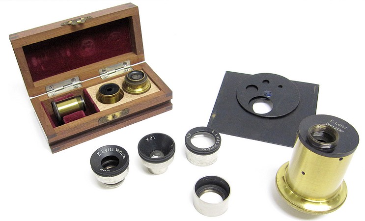
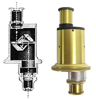
The erecting prism attachment utilizes two Porro-prisms that are mounted within the tube in such manner that, by two successive reflections of the image formed by the objective, an erect image is obtained in the eyepiece, which is of the Ramsden type. Included are two objectives for use with the erector and three objectives for use as a conventional dissecting microscope. The light path through the optics is illustrated on the left.
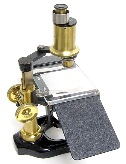
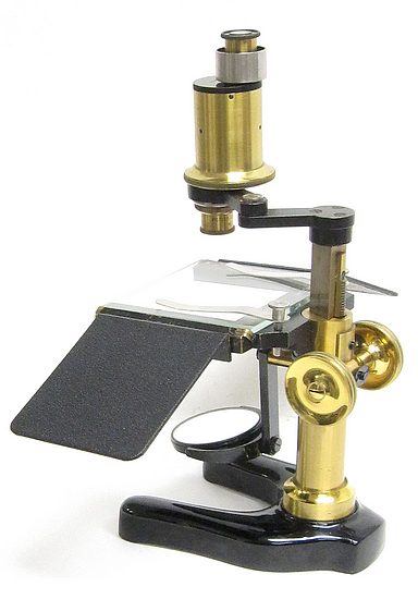
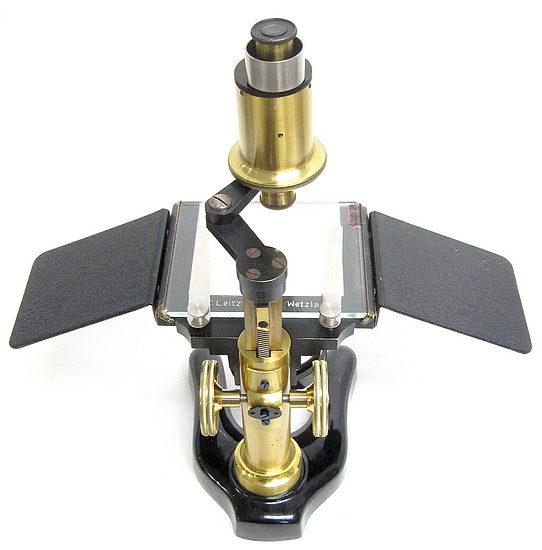
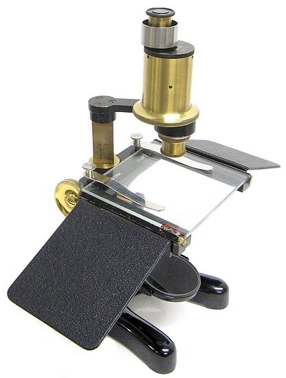
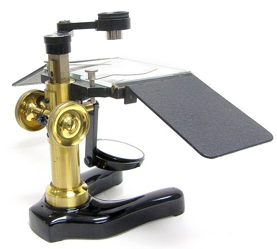
Subsequently, this erecting apparatus was supplied with other Leitz dissection stands such as the model shown on this page. It is described in the 1929 catalog as the "Dissecting Microscope Model W".
From the 1929 Leitz catalog:
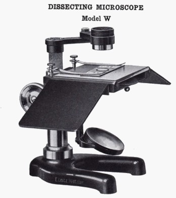
Focusing Adjustment — By rack and pinion with milled head on either side, giving a range of 80mm.
Stage Glass — 80 x 95mm, removable; second set of grooves beneath stage accommodate a metal plate , with circular disc to provide white or black background; spring clips attached to stage support.
Hand Rests — Of metal, 90mm in length and detachable, leather covered.
Base and Pillar — Base of metal, modified horseshoe form; round pillar, of metal, attached to base.
Mirror — Plano 50mm diameter, one side of opal glass; mounted on swinging arm.
Lens — “Doublet” or triple “Aplanat” are regularly supplied; lens arm will hold any of our regular magnifiers for dissecting microscopes
Finish — Base in black alcohol proof lacquer, pillar in blond lacquer.
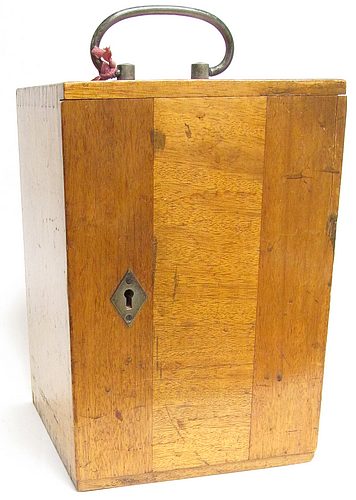
Case — Of wood, highly polished and fitted with lock and key.

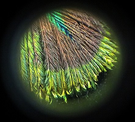
A view through the microscope
(peacock feather)
