The following was extracted from: Clinical Hematology by J.C. DaCosta, 1902
SPECTROSCOPICAL EXAMINATION.
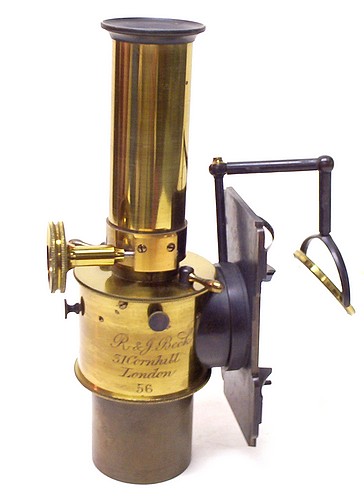
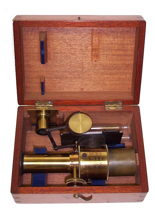
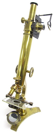
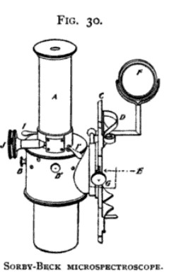
For clinical work the Sorby-Beck microspectroscope, to be used in connection with the microscope, is an excellent instrument, being both accurate, and, comparatively speaking, easy to manipulate. Other very perfect instruments for the spectroscopical examination of the blood, differing but little from the original Sorby model, are Fig. 30. also made by Zeiss, by Leitz, and by Browning.
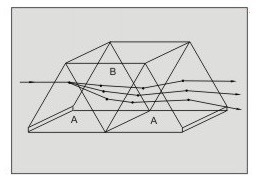
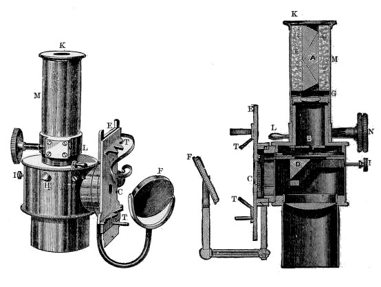
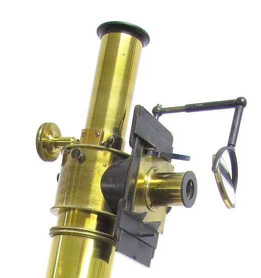
This instrument (Fig.The 30) when in use fits into ocular, for which it is substituted. Its essential part consists of a tube, A, in which a series of five prisms, two of flint and three of crown glass, is arranged in such a manner that the emergent rays, which are separated by dispersion, leave the prisms in practically the same direction as that taken by the entering immergent ray. At one side of the tube is fixed a right-angle reflecting prism, so that the spectrum of a solution of normal blood may be thrown alongside that of the specimen under investigation, the two spectra thus being comparable. The adjustment of the spectra is effected by means of the two small screws, B, B'. The receptacle containing the control solution of blood is clamped to the stage, C, by a spring clip, D, light being reflected through the liquid and into the rectangular aperture, E, by the swinging mirror, F. The width of this aperture is controlled by the screw, G. The receptacle containing the blood solution to be examined is placed upon the stage of the microscope, being brought into focus with a low-power (2/3 or 1 inch) dry objective. Beneath the tube enclosing the series of prisms is mounted an achromatic ocular, below which a narrow slit-like diaphragm is situated, the vertical size of this opening being regulated by a milled screw, not shown in the illustration, and its breadth by the two small levers, /, /'. Both ocular and prisms may be moved simultaneously toward and away from the diaphragm, by a rack-and-pinion mechanism controlled by the wheel, J, so that any part of the spectrum may be brought into focus.
The liquids to be examined should be placed in Sorby's tubular cells, and cover-glasses superimposed. These cells (Fig. 31) are narrow lumened glass receptacles made of barometer tubing, both ends of which are accurately ground to parallel surfaces, one end being cemented to a small polished glass plate.
Method of Examination. The specimen of blood obtained in the usual manner, by puncture, is first diluted with distilled water one hundred times, by means of the Thoma-Zeiss erythrocytometer, and sufficient of this laked blood dropped into a Sorby cell to fill it exactly to the brim. A cover-glass is then carefully laid over the open end of the cell, the precaution being taken to prevent the formation of air-bubbles upon the surface of the column of liquid thus enclosed. A second cell, to be used as the control, is filled with normal blood, similarly diluted, and both are then adjusted in their respective positions, as already explained.
In making the examination, a ray of artificial light (that from a Welsbach incandescent burner being most suitable) is projected by the microscope mirror through the lumen of the cell containing the suspected blood, and the surface of the liquid focused with an ordinary ocular. The latter is then removed from the microscope tube and replaced by the spectroscope ocular, and the second spectrum, that of the normal blood, is brought into proper position alongside that of the first, so that any differences between the two may be contrasted by the observer.
The appearance of the spectra of normal and of pathological blood, together with the circumstances under which the latter occur, have been described in another section.
Also, see the description of the microspectroscope in the 1888 Beck catalog.