
From The Journal of the Royal Microscopical Society, 1902, page 353
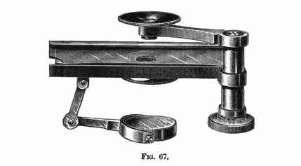
Pillischer's "Lenticular Microscope" Mr. J. Pillischer, of Bona Street, has most kindly presented this very interesting portable, really pocket, Microscope to the Society's Cabinet. It was designed by the late Mr. M. Pillischer, the donor's uncle.
The instrument is figured and described in Urinary Deposits by Golding Bird (p. 29, fig. 13, 1857, 5th ed.), but it will be noticed that the figure differs slightly from the original, inasmuch as a second spring to hold the slide has been added, and a semicircular segment cut out at both ends instead of at one end of the base-plate as there shown.
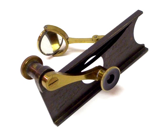
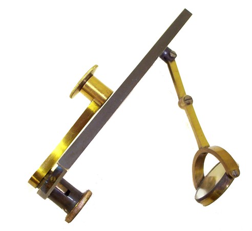
The design of this instrument (fig. 67) is most ingenious: there is neither stand nor limb, the main basis of the instrument being the slide- holder, at one angle of which is a short pillar containing a direct-acting screw fine adjustment, which acts upon a swinging arm carrying the lens. Below the stage is a mirror attached to a jointed arm, and a wheel of diaphragms. The lenses, three in number, are Coddingtons of 1/4, 1/10 and 1/25 -in. foci.
It may be pointed out, says E. M. Nelson, that an instrument of this kind, fitted with achromatic loups, would be very serviceable to a microscopist for field work.
It will be remembered that three of Dr. Gairdner's Microscopes, made by Bryson of Edinburgh, were exhibited, figured, and described in the journal for 1899, p. 643, fig. 149.
These had Coddington lenses, each power having a separate Microscope to itself. Gairdner's Microscope was described in the first edition of Carpenter on the Microscope, 1866, p. 74, fig. 15, and there it is said to be of use in bed-side investigations of urinary deposits.
In design, Gairdner's Microscope is far inferior to that of Pillischer's, inasmuch as there is no possibility of either moving the slide under the lens, or the lens over the slide, so nothing can be seen except the single point iu the axis of the lens.
From: Urinary Deposits by Golding Bird, 6th Edition, 1863
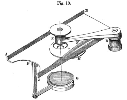
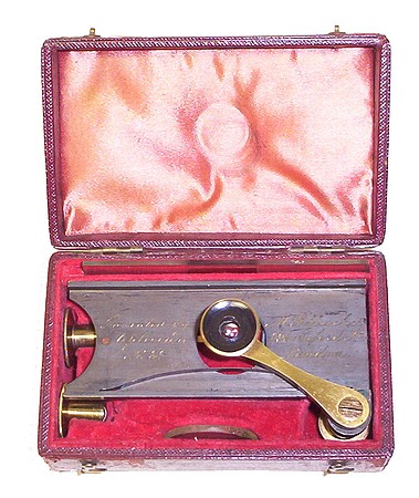
It has often been a matter of regret that a very portable microscope has not hitherto been contrived, sufficiently small to be easily carried in the pocket, and sufficiently economical to be within the reach of all. Very recently Mr. Pellischer has constructed one, which he terms the "lenticular microscope," which seems to me to fulfil this condition entirely, and I cannot too strongly recommend it to the notice of the profession. In the construction of this beautiful little instrument, he has made use of the excellent and well-known Coddington lens, which consists of a very thick double convex lens excavated at the sides into a kind of dumb-bell shape, by which the extreme lateral rays are cut off, and a very perfect image obtained. It consists of a rectangular piece of brass (ABC D), excavated at one end, furnished with raised sides. To the inner surface of the side C D, a steel spring (E) is fixed for the purpose of keeping the piece of glass on which the object is placed quite steady. At the under surface at F a brass arm is fixed bearing a small concave mirror (G). A perforated diaphragm (H) is fixed to a pin beneath an aperture in the plate, so that by moving it, the pencil of rays reflected from the mirror may be lessened, and a clearer definition obtained. D K is a strong arm of brass, capable of being moved horizontally over the aperture in the plate, whilst a fine screw movement at D enables it to be raised vertically. The lenses, having respectively a focal length of about 1/4, 1/20, and 1/35th of an inch each, are placed in a split cylinder (K) at the end of the arm (D K). When not in use the arm to which the mirror is attached is folded up flat against the under surface of the plate, and thus the whole apparatus can be carried in the waistcoat-pocket. To use this instrument, a drop of urine containing a deposit is placed on a slip of glass, and covered with a piece of mica or thin glass. It is then placed on the plate (ABC D), on which the spring firmly retains it. One of the lenses being then placed in the cylinder (K), the object is brought into focus by means of the screw (D), illumination being effected by holding up the instrument to the light of the clouds or a candle, or still better by reflecting a ray of light through the object by means of the mirror. If the object is very translucent, especially when epithelial cells are searched for, the amount of transmitted light should be diminished by means of the diaphragm. Should the deposit consist of large coarse crystals, it is better placed in a little cavity ground in a plate of glass (which accompanies the instrument), as they will thus escape injury when cover with a piece of thin glass for examination.
This very early version (serial number 32) predates the incorporation of the perforated diaphragm wheel (H). The set is complete with three objectives and a glass disk.
Moritz Pillischer established his business as a scientific instrument maker around the 1840's. He made or sold a variety of scientific instruments and is well known for his microscopes. He retired in 1887 and the business was taken over by his nephew, Jacob Pilliischer. The firm continued well into the 20th century. Pillischer was located at the address marked on this instrument during the years 1851 to 1853.See the Pillischer Family Timeline. Also, see this detailed essay on the Pillischer business.
An 1854 advertisement